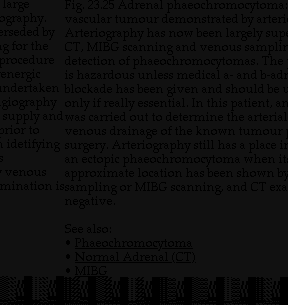
Labels:text | screenshot | black and white | black | font | document OCR: Large Fig. 23.25 Adrenal phaeochromocytoma ography. vascular tumour demonstrated by arteri rseded by g for the Arteriography has now been largely sup CT, MIBG scanning and venous samplin procedure detection of phaeochromocytomas. The is hazardous unless medical a- and b-ad undertaken blockade has been given and should be u energic giography only if really essential. In this patient, ar supply and was carried out to determine the arterial prior to venous drainage of the known tumour idetifying surgery. Arteriography still has a place in an ectopic phaeochromocytoma when it venous approximate location has been shown by mination issampling or MIBG scanning, and CT exa negative. See also: · Phaeochromocytoma · Normal Adrenal (CT) . MIBG